Cell Mechanics
In this section, I will show the key points of one my published cell mechanics studies. Since in this study, I use a lot of techniques that I describe in other sections, I will simply refer to these without re-describing them.
Stress Fiber Laser Nanosurgery
 Fig. 1. Time course of laser nanosurgery of central and peripheral stress fibers (SFs)
Traction forces against the extracellular matrix are crucial to cellular motility and mechanosensing. They can be generated by stress fibers (SFs), which are bundles of filamentous actin, actin-binding proteins, and non-muscle myosin II. To study SF viscoelastic properties, we applied single SF laser nanosurgery in different sub-populations (central and peripheral SFs), and concluded that they have different retraction dynamics.
Some SF laser nanosurgury examples are shown in Fig. 1. As contractile structures, SFs retracted in short time scale (in seconds) following laser nanosurgery, which was achieved by high energy deposition from a femtosecond pulsed laser of a confocal / two-photon microscopy system. (Reference: Chang, C. W., Kumar, S., Journal of Cell Science, 2013, 126(14), 3021-3030)
Stress Fiber Structural Model
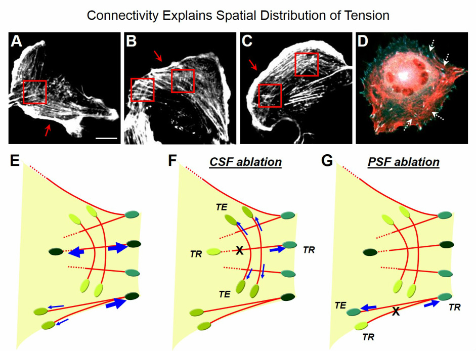 Fig. 2. Structural model of SF network demonstrating how the inter-connectivity of central SFs helps force distribution
Using the FRET-based vinculin tension sensor (VinTS; see the FRET section) combined with laser nanosurgery, followed by image analysis on the correlation between focal adhesion (FA) tension change and orientation (see the Image Analysis section), we reached the model shown in Fig. 2. In our model, the interconnected central SFs (red boxes, Fig. 2A-C) re-direct tension to a broader array of FAs (white dotted arrows, Fig. 2D) with a diversity of cellular locations and orientations (Fig. 2F), which preserves cell shape after SF nanosurgery. Conversely, peripheral SFs, which are not as structurally coupled into the total SF network (red arrows, Fig. 2A-C), distribute dissipated tension to a narrower segment of cellular FAs with similar orientations (Fig. 2G). Thus, peripheral SF disruption is more likely to trigger FA rupture and cellular contraction. In Fig. 2E-G, green ovals represent FAs, with a darker green color indicating higher tension in that FA; blue arrows represent force vectors along SFs, with stronger force reflected by thicker arrows; red solid or dashed lines represent SFs; the laser nanosurgery site is indicated by an “X”; "TE" indicates tension-enhanced FAs; "TR" indicates tension-reduced FAs. (Reference: Chang, C. W., Kumar, S., Journal of Cell Science, 2013, 126(14), 3021-3030)
